Dataset
Research Foundation
Using spike data recorded from multiple brain regions while subjects observe urban scenes, we apply dimensional reduction techniques (PCA, UMAP) and topological data analysis to uncover region-specific manifolds. These manifolds reorganize visual complexity into distinct geometric structures that reveal the brain's computational principles.
Experimental Environment
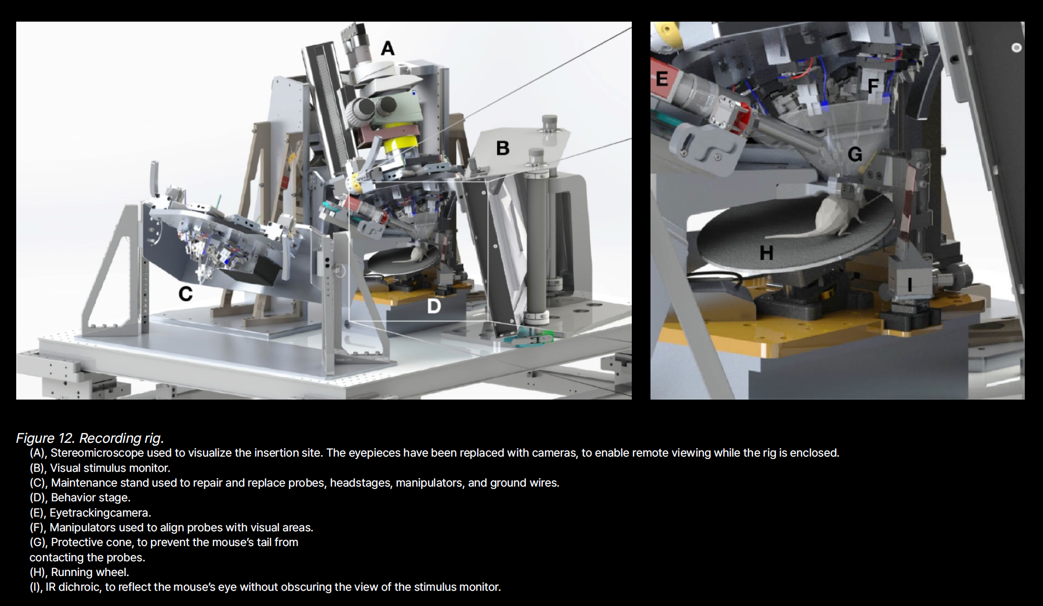
Experimental setup for neural recording during visual stimulation
Visual Stimuli
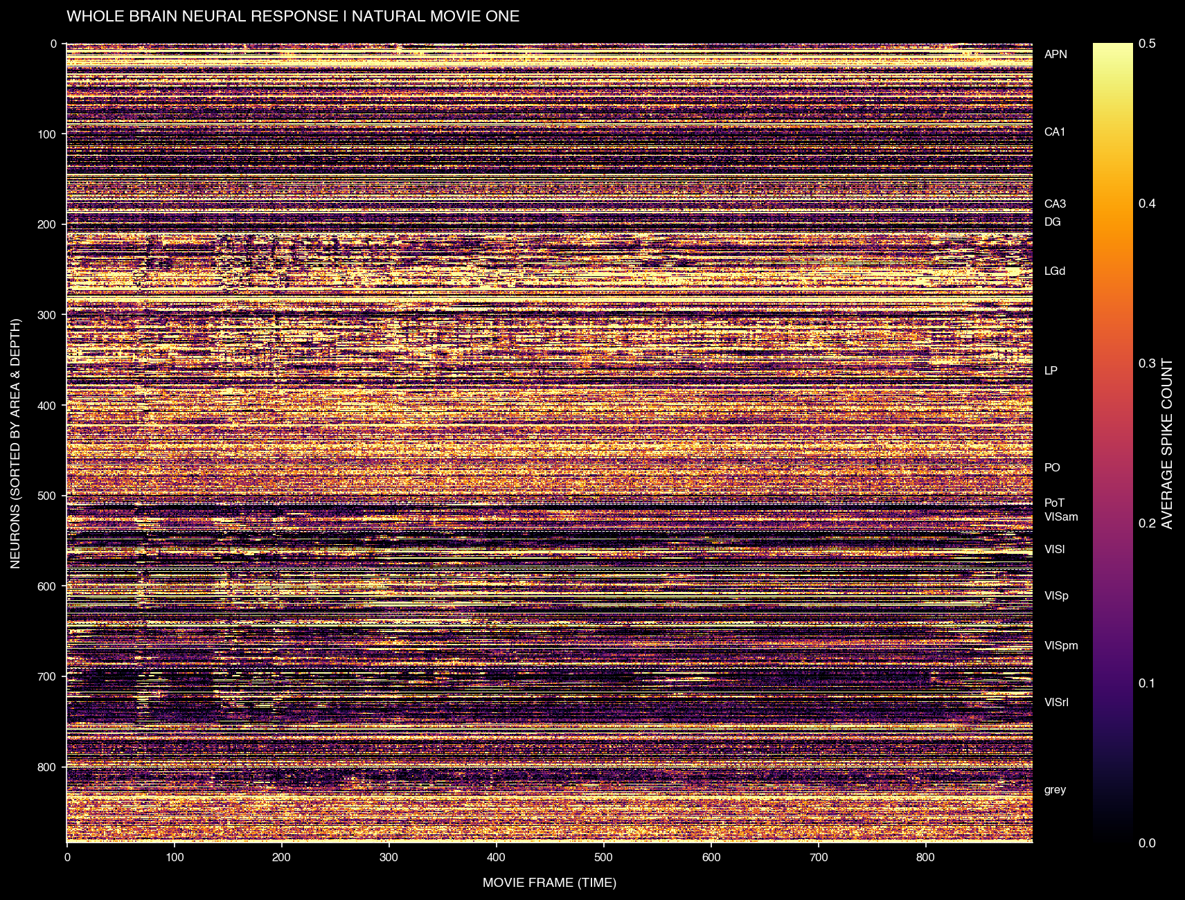
Natural movie stimulus presented to subjects during neural recording
Neural Data & Recording
Neuropixels Probes — High-density silicon electrodes capable of recording from hundreds of neurons simultaneously across multiple brain regions. Each probe contains 384 recording sites distributed along a 10mm shaft, allowing researchers to capture neural activity from different cortical layers and subcortical structures.
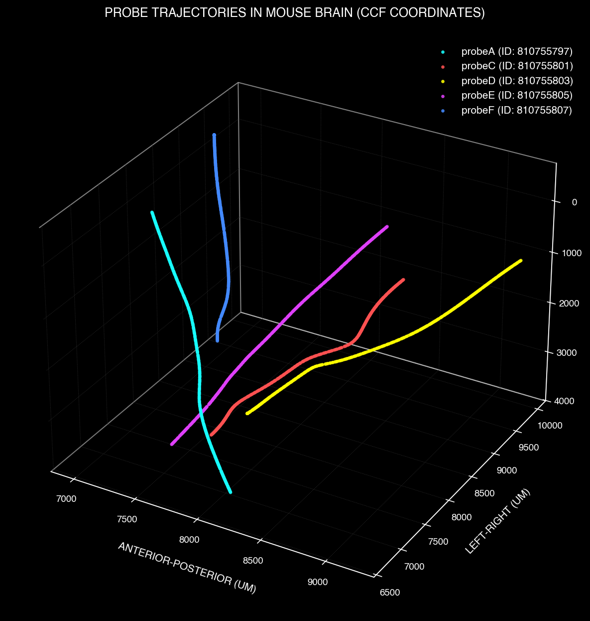
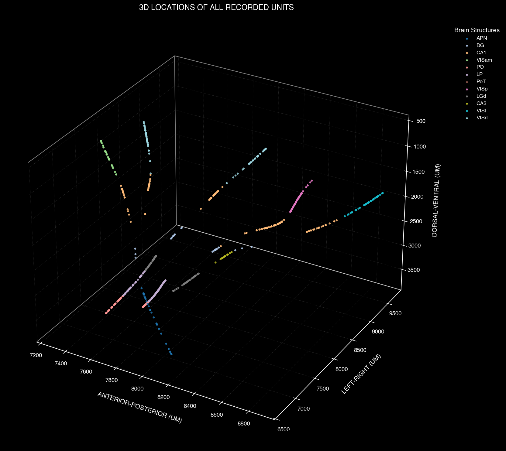
Neuropixels probe configuration and brain region mapping
Brain Regions Analyzed:
- Visual Cortex (VISp, VISl, VISam, VISpm, VISrl) — Primary and higher-order visual processing areas
- Thalamus (LP, LGd, PO, VPM) — Visual relay stations and sensory integration
- Hippocampus (CA1, DG) — Memory formation and spatial representation
LFP Signals
Raw Local Field Potential (LFP) signals from neural recordings
Analytical Pipeline
From Raw Data to Geometry: The analysis transforms high-dimensional neural spike data through multiple stages to reveal intrinsic geometric structure.
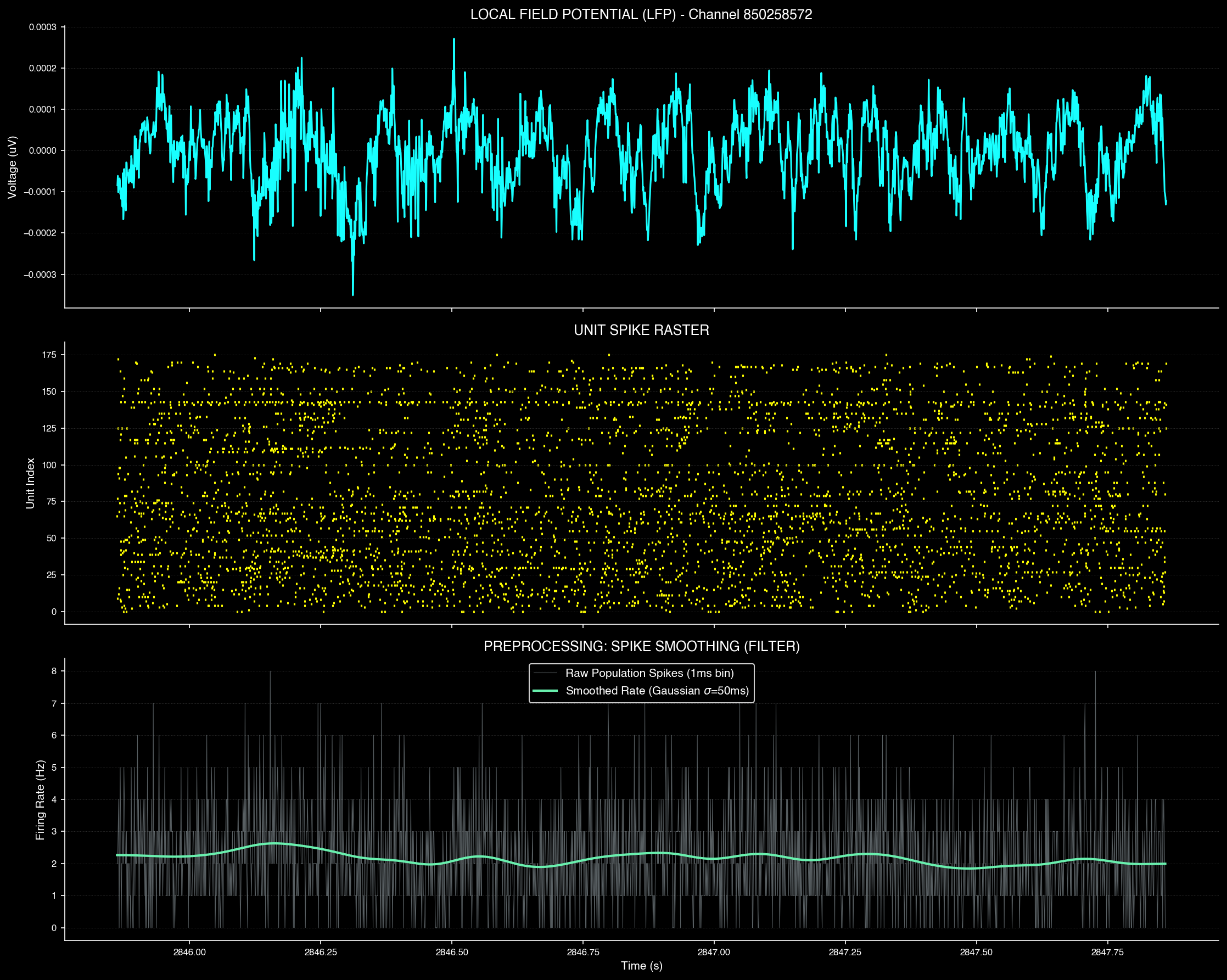
Spike data preprocessing and filtering
Next Steps
To understand how this raw neural data is transformed into interpretable geometric structures, explore the Topology section where we detail the complete analytical pipeline from dimensionality reduction to topological analysis.